How Does It Help?
Why Choose the Echolight REMS Scan?
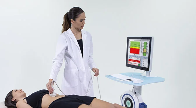
The Echolight REMS Scan helps by providing a non-radiation based measurement tool that is low cost, low risk and time effective. Once a baseline measurement has been established, it is recommended that you have your scan repeated in approximately 12 months to assess the effectiveness of any interventions that you have partaken in to inform decisions about how to manage this in the future. This will be guided by your clinician and is tailored to what you need and how it can fit into your lifestyle.
What Does REMS Measure?
REMS technology scans the spine and femur, the two areas most prone to osteoporotic fracture using ultrasound signals to produce two key measurements:
Bone mineral density
This is scored two ways: a T-score (how your bones compare to a healthy young adult) and a Z-score (how they compare to others your age). A T-score below –2.5 indicates osteoporosis; a score between –1 and –2.5 indicates osteopenia.
Fragility Score
This measures the quality and structural integrity of your bone. It’s a unique marker of bone microarchitecture and research shows it is, in fact, a more reliable predictor of fracture risk than density alone. In other words, two people with identical T-scores can have very different real-world risk of fracture.
Together, these two measurements give one of the most accurate and complete pictures of bone health.
Echolight REMS vs DEXA Scan
Both REMS and DEXA measure bone mineral density and produce a T-score. The key differences are:
Echolight REMS | DEXA Scan | |
Type of Radiation | Ultrasound (radiation-free, safer for more frequent use) | Low-level X-ray |
Fragility Score | “Tick symbol” | x |
Objectivity | Computer-generated reading, reduces variability of human interpretation | Radiologist interpretation |
Accessibility | Available here without external referral, with immediate results | Referral required |
For many patients, REMS is a highly effective alternative to DEXA. For those who have already had a DEXA scan, it provides complementary information that builds a richer picture of bone health.
FAQs
Frequently Asked Questions
What is the difference between REMS and DEXA?
Both measure bone mineral density and produce a T-score, which is used to diagnose osteoporosis (T-score below –2.5) or osteopenia (T-score between –1 and –2.5). REMS differs in three key ways: it uses ultrasound rather than radiation; it produces a Fragility Score — a measure of bone microarchitecture shown in research to be more accurate in predicting fracture risk; and it generates a computer-automated reading, reducing the variability associated with radiologist-interpreted DEXA scans.
At what age should I start thinking about bone health?
It is never too early or too late. In your teens and twenties, the focus is on building peak bone mass — achieved through sport, strength training and good nutrition. Peak bone mass is reached in the mid-twenties, and the stronger your bones at that point, the longer before age-related decline becomes clinically significant. From your mid-thirties onwards, and particularly around menopause, proactive monitoring and intervention become increasingly important. For anyone with risk factors — family history, low body weight, certain medications — earlier assessment is advisable regardless of age.
Can exercise reverse low bone density?
Yes. A landmark Australian trial (2018) demonstrated that participants who undertook supervised strength training twice weekly for nine months — alongside a weekly BioDensity session — showed improvements in bone mineral density compared to those who performed bodyweight exercise, balance training and stretching. The Royal Osteoporosis Society recommends strength training as a first-line intervention. At Mayfair Health, our bone health programme is designed specifically around this evidence.
Is it safe to exercise with osteoporosis or low bone density?
Not only is it safe — it is necessary. Bone formation is stimulated by mechanical loading, and exercise is the cornerstone of our bone health programme. Our physiotherapists are trained to safely and progressively load the skeleton through supervised strength work, using machines and weights to stimulate an adaptive bone response. In the Australian trial referenced above, no significant injuries were reported despite participants lifting heavy relative to their baseline strength.
Should high-impact activity be avoided?
Not necessarily. High-impact activity stimulates bone adaptation and is a valuable component of a bone health programme for many patients. Whether it is appropriate depends on individual factors — including the Fragility Score from the REMS scan, which measures bone quality and can help determine whether a patient is suitable for higher-impact loading. Your clinician will advise based on your specific results.
Why are women more at risk of osteoporosis?
Oestrogen regulates bone formation, and during menopause oestrogen levels fall significantly — slowing bone formation while bone resorption continues, leading to a net loss of bone mineral density over time. Women who experience early or surgical menopause, irregular periods or certain gynaecological conditions are at increased risk and should consider earlier assessment. Research shows that 1 in 2 women over 50 will develop osteopenia or osteoporosis — making proactive screening particularly important in this group.
Are there any signs or symptoms of low bone density?
Osteoporosis and low bone mineral density are known as silent conditions — there are no symptoms until a fracture occurs. For many people, a fracture is the first indication that their bones have been weakening for years. This is precisely why proactive scanning is so valuable: identifying bone loss early allows meaningful intervention before significant deterioration occurs.
How often should I have the scan repeated?
We recommend repeating the scan at approximately 12 months following your initial scan to assess the effectiveness of any interventions. Frequency thereafter is guided by your clinician based on your results, risk profile and the programme you are undertaking.
Is the scan covered by private health insurance?
This varies between insurers and policies. We recommend contacting your insurer in advance to confirm your level of cover. Our team is happy to help with any queries.